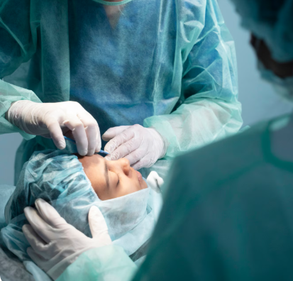
Treatment Overview
Fovea-Sparing Retinal Detachment Surgery is a precision retinal procedure designed to repair macula-on retinal detachments while preserving the fovea, the central region of the retina responsible for sharp central vision. This technique is particularly important for patients whose detachment threatens the macula but where the fovea remains attached. By carefully manipulating the detached retina, surgeons can reattach the peripheral retina without compromising central vision.
In Korea, this surgery is performed using state-of-the-art technology and specialized surgical expertise:
- 25–27G microincision pars plana vitrectomy (PPV) for minimal trauma
- Intraoperative OCT (iOCT) for real-time monitoring of foveal position
- Widefield fundus visualization to map the detachment and tears
- Endolaser photocoagulation or diathermy for peripheral retinal break sealing
- AI-assisted surgical planning to preserve the fovea during reattachment
- Combination with gas or silicone oil tamponade, cryopexy, or scleral buckle if necessary
This approach allows surgeons to reattach the retina safely, reduce the risk of foveal damage, and optimize long-term visual outcomes.
Purpose & Benefits
Purpose
- Repair macula-on retinal detachments while preserving foveal function
- Flatten detached peripheral retina and seal retinal breaks
- Minimize traction on the fovea to protect central vision
- Complement adjunctive procedures such as tamponade, laser, or cryopexy
- Prevent progression to total retinal detachment
- Restore retinal anatomy while optimizing functional outcomes
Benefits
- Preserves sharp central vision by avoiding foveal manipulation
- Minimally invasive with microincision vitrectomy
- High anatomical and functional success rates in macula-on detachments
- Can be combined with gas, silicone oil, laser, or cryopexy for optimal results
- Real-time intraoperative OCT enhances surgical precision
- Rapid postoperative recovery with minimal complications
- Ideal for high-risk patients where foveal preservation is critical
Ideal Candidates
Fovea-Sparing Retinal Detachment Surgery in Korea is ideal for:
- Patients with macula-on retinal detachment
- Eyes with peripheral retinal tears or holes threatening the fovea
- Individuals at risk of foveal involvement during standard retinal repair
- Patients requiring adjunctive endolaser, diathermy, or cryopexy
- Trauma-induced retinal detachments where macula remains attached
- High myopia patients with macula-on detachments
- Patients undergoing pars plana vitrectomy for complex detachments
Possible Risks & Complications
This surgery is highly precise but carries some potential risks:
Minor/Transient Issues
- Temporary blurred vision
- Mild eye irritation or discomfort
- Small floaters due to residual vitreous
Rare/Serious Risks
- Accidental foveal detachment or damage
- Recurrent retinal detachment
- Retinal or choroidal hemorrhage
- Cataract progression in phakic eyes
- Endophthalmitis (very rare)
- Tamponade-related visual distortion or scotomas
Korean clinics minimize risks through:
- Intraoperative OCT for real-time foveal monitoring
- AI-assisted mapping of retinal tears and detachment edges
- Microincision vitrectomy to reduce traction on the macula
- Experienced retinal surgeons specialized in fovea-sparing techniques
- Strict postoperative monitoring with multimodal imaging
Related Diagnostic & Treatment Techniques
- Pars Plana Vitrectomy (PPV) – Standard microincision vitrectomy
- Endolaser Photocoagulation – Sealing peripheral retinal breaks
- Diathermy or Cryopexy – Adjunctive retinal repair methods
- Gas or Silicone Oil Tamponade – Maintains retinal reattachment
- Intraoperative OCT (iOCT) – Real-time foveal and retinal monitoring
- Widefield Fundus Imaging & OCT – Pre- and post-surgery assessment
- AI-Assisted Surgical Planning – Optimizes fovea-sparing technique
Treatment Process in Korea
Step 1 – Preoperative Assessment
- Comprehensive eye exam including visual acuity and IOP
- OCT and widefield fundus imaging to assess macula and detachment
- AI-assisted analysis to plan surgical strategy while preserving fovea
- Mapping of retinal breaks and peripheral detachment zones
Step 2 – Surgical Procedure
- Local or general anesthesia
- 25–27G microincision vitrectomy performed
- Careful removal of vitreous traction near the fovea
- Peripheral retinal tears sealed with endolaser, diathermy, or cryopexy
- Controlled fluid–air or fluid–gas exchange to flatten retina
- Gas or silicone oil tamponade applied if necessary
Step 3 – Postoperative Follow-Up
- Immediate postoperative examination within 24 hours
- Follow-up at 1 week, 1 month, and 3 months
- OCT and fundus imaging to confirm retinal reattachment and foveal integrity
Duration: 60–120 minutes
Setting: Advanced retinal surgery operating room
Recovery & After-Care
After-Care Guidelines
- Use prescribed antibiotic and anti-inflammatory eye drops
- Maintain head positioning if gas tamponade is used
- Avoid heavy lifting, bending, or strenuous activity for several weeks
- Protect the eye from trauma and contaminants
- Attend all scheduled postoperative appointments
Recovery Timeline
- Immediate: Mild blurred vision, especially with gas tamponade
- 1–2 Weeks: Retina stabilizes; initial visual improvement
- 1–3 Months: Vision improves as retinal adhesion solidifies
- 3–6 Months: Long-term foveal preservation and retinal stability confirmed
Results & Longevity
Expected Results
- Successful reattachment of detached peripheral retina
- Preservation of foveal structure and central vision
- Reduced risk of recurrent detachment
- Can be combined with adjunctive procedures for optimal outcomes
- High anatomical and functional success rates
Longevity
- Permanent retinal attachment achieved in most cases
- Lifelong monitoring recommended for high-risk patients
- Additional intervention rarely needed if procedure is successful
Why Korea Is a Top Destination
- Advanced microincision vitrectomy and fovea-sparing techniques
- Intraoperative OCT for real-time macular monitoring
- AI-assisted surgical planning for precision and safety
- Experienced retinal surgeons specializing in macula-on detachments
- Integration with laser, diathermy, cryopexy, and tamponade for optimal outcomes
- Multimodal imaging and efficient outpatient care
- English-friendly clinics for international patients
Unique Korean Innovations
- AI-guided mapping of retinal breaks to protect fovea
- Real-time iOCT monitoring for macula-on retinal detachments
- Microincision vitrectomy for minimal macular traction
- Controlled fluid–air/gas exchange to avoid foveal detachment
- Hybrid approaches combining laser, diathermy, and tamponade
- Digital surgical dashboards for postoperative retinal and foveal tracking
Cost Range (Indicative Estimate)
| Package | Price (KRW) | Approx. USD | Inclusions |
|---|---|---|---|
| Standard Fovea-Sparing PPV | ₩6,500,000 – ₩10,000,000 | ~$5,000 – $7,650 | Microincision PPV + laser/diathermy + imaging |
| Fovea-Sparing PPV + Gas Tamponade | ₩7,000,000 – ₩10,500,000 | ~$5,350 – $8,000 | PPV + gas + adjunct retinal repair |
| Fovea-Sparing PPV + Silicone Oil Tamponade | ₩8,000,000 – ₩12,000,000 | ~$6,150 – $9,200 | PPV + silicone oil + laser/diathermy |
| Postoperative Monitoring Package | ₩300,000 – ₩800,000 | ~$230 – $620 | OCT + fundus + AI-assisted follow-up |
Popular Clinics in Korea
- Kim’s Eye Hospital (Seoul)
- Gangnam Severance Hospital Retina Unit
- Seoul National University Hospital Retina Center
- B&VIIT Eye Center (Seoul)
- BGN Eye Clinic (Seoul & Busan)
- NUNE Eye Hospital (Daegu)
- Dream Eye Center (Seoul)
- Glory Seoul Eye Clinic


