Treatment Overview
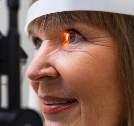
Laser Retinopexy is a precise, minimally invasive laser procedure used to treat peripheral retinal tears or holes before they progress to retinal detachment. The treatment involves creating circumferential laser burns around the tear, which induce a strong adhesive scar between the retina and underlying tissue, stabilizing the retina and preventing fluid accumulation.
In Korea, this procedure is performed with state-of-the-art retinal laser systems and highly skilled ophthalmologists:
- Focal or pattern laser photocoagulation targeting the retinal tear and surrounding tissue
- Widefield retinal imaging for accurate localization of peripheral tears
- High-resolution OCT and fundus photography to evaluate retinal health pre- and post-procedure
- AI-assisted laser planning to optimize spot placement and energy settings
- Rapid outpatient procedure with minimal discomfort and no incisions
Laser retinopexy is highly effective for preventing retinal detachment in patients with peripheral tears, lattice degeneration, or high-risk retinal areas.
Purpose & Benefits
Purpose
- Seal peripheral retinal tears and prevent progression to detachment
- Strengthen retinal adhesion in high-risk areas
- Treat lattice degeneration or symptomatic peripheral retinal holes
- Reduce the need for surgical intervention in early-stage retinal disease
- Complement ongoing monitoring in patients with vitreoretinal disease
Benefits
- Minimally invasive with no incisions
- Quick outpatient procedure (typically <30 minutes)
- Prevents retinal detachment in high-risk patients
- Precise targeting using widefield imaging and AI-assisted planning
- Can be combined with OCT, fundus photography, and fluorescein angiography for comprehensive assessment
- Minimal discomfort and rapid recovery
- Highly effective preventive retinal treatment
Ideal Candidates
Laser Retinopexy for Peripheral Retinal Tears in Korea is ideal for:
- Patients with symptomatic or asymptomatic peripheral retinal tears
- Individuals with lattice degeneration or thinning of the peripheral retina
- High myopia patients at risk for retinal detachment
- History of retinal tear or prior retinal detachment in one eye
- Patients with vitreous traction on the peripheral retina
- Preemptive treatment before cataract surgery in high-risk eyes
Possible Risks & Complications
Laser retinopexy is generally safe, but potential risks include:
Minor/Transient Issues
- Mild eye discomfort during or after laser
- Temporary blurred vision or light sensitivity
- Small scotomas at laser spots
Rare/Serious Risks
- Retinal burn or damage to adjacent tissue
- Mild localized hemorrhage at laser spots
- Induction of new retinal breaks (very rare)
- Rare inflammation or infection
Korean clinics minimize risks through:
- High-resolution imaging and widefield mapping for precise targeting
- AI-assisted laser energy calibration
- Experienced retinal specialists performing the procedure
- Post-treatment OCT/fundus confirmation of laser effect
- Immediate follow-up care to address any complications
Related Diagnostic & Treatment Techniques
- OCT (Optical Coherence Tomography) – Evaluates retinal structure
- Fundus Photography & Widefield Imaging – Maps peripheral retinal tears
- Fluorescein Angiography (FA) – Assesses perfusion and leakage if needed
- Pattern or Focal Laser Photocoagulation – Creates retinal adhesion around tears
- Prophylactic Retinal Surgery – For complex or recurrent detachments
Treatment Process in Korea
Step 1 – Preoperative Assessment
- Comprehensive eye exam including visual acuity, IOP, and fundus evaluation
- OCT and widefield fundus imaging to locate peripheral retinal tears
- AI-assisted planning for laser spot placement and energy adjustment
Step 2 – Laser Procedure
- Topical anesthesia applied to the eye
- Patient positioned at the retinal laser system
- Laser delivered in a circumferential pattern around the peripheral tear
- Retinal whitening observed to confirm effective adhesion
- Multiple spots applied if necessary, guided by imaging
Step 3 – Post-Treatment Follow-Up
- Immediate examination to check retinal integrity
- Follow-up at 1 week and 1 month with OCT or fundus imaging
- Monitoring for new tears or detachment risk
Duration: 15–30 minutes per session
Setting: Outpatient retinal clinic
Recovery & After-Care
After-Care Guidelines
- Resume normal activities immediately
- Avoid rubbing the treated eye
- Use prescribed anti-inflammatory eye drops if recommended
- Attend scheduled follow-up imaging sessions
- Monitor for new symptoms such as flashes, floaters, or vision changes
Recovery Timeline
- Immediate: Vision may be mildly blurred from laser effect
- 1–2 Days: Retinal adhesion begins to stabilize
- 1–4 Weeks: Barricade adhesion fully established
- Long-Term: Maintains retinal stability and prevents detachment
Results & Longevity
Expected Results
- Successful sealing of peripheral retinal tears
- Prevention of progression to retinal detachment
- Long-term stabilization of high-risk retinal areas
- Reduced likelihood of surgical intervention
- Can be combined with OCT and follow-up imaging for ongoing monitoring
Longevity
- Lifelong retinal stability expected if no new tears develop
- Periodic monitoring recommended, especially in high-risk eyes
- Repeat laser therapy may be applied if new tears appear
Why Korea Is a Top Destination
- Advanced retinal laser systems with widefield imaging
- AI-assisted planning for precise laser delivery
- Experienced retinal specialists performing preventive procedures
- Integration with OCT, fundus photography, and fluorescein angiography
- Rapid outpatient treatment with minimal discomfort
- Personalized follow-up for long-term retinal stability
- English-friendly clinics for international patients
Unique Korean Innovations
- AI-assisted laser energy calibration for optimal adhesion
- Widefield and OCT-guided retinal mapping
- Digital dashboards for longitudinal retinal monitoring
- Combination protocols with preventive OCT and fluorescein imaging
- Streamlined outpatient laser services with rapid recovery
Cost Range (Indicative Estimate)
| Package | Price (KRW) | Approx. USD | Inclusions |
|---|---|---|---|
| Single Laser Retinopexy Session | ₩200,000 – ₩400,000 | ~$150 – $310 | Laser photocoagulation around peripheral retinal tear + imaging |
| Comprehensive Assessment + Laser | ₩500,000 – ₩900,000 | ~$380 – $700 | OCT + fundus + laser + AI-assisted planning |
| Follow-Up Monitoring Package | ₩300,000 – ₩800,000 | ~$230 – $620 | 1–3 follow-up imaging sessions + evaluation |
Popular Clinics in Korea
- Kim’s Eye Hospital (Seoul)
- B&VIIT Eye Center (Seoul)
- BGN Eye Clinic (Seoul & Busan)
- Gangnam Severance Hospital Retina Unit
- Seoul National University Hospital Retina Center
- Dream Eye Center (Seoul)
- NUNE Eye Hospital (Daegu)
- Glory Seoul Eye Clinic


