Treatment Overview
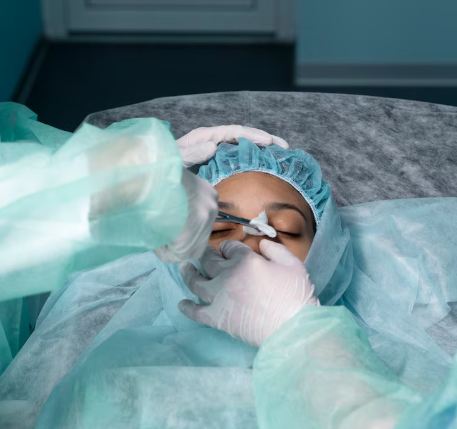
Anatomic Retinal Reattachment Surgery is a specialized procedure aimed at restoring the normal anatomical position of the retina after it detaches from the underlying tissue. This surgery is essential to prevent permanent vision loss and is performed using advanced techniques that ensure precise reattachment, restoration of retinal layers, and stabilization of the eye structure.
In Korea, this treatment is performed using world-leading retinal surgical systems and modern imaging technologies such as:
- Microincision pars plana vitrectomy (25G–27G) for minimal trauma
- Scleral buckling techniques for external retinal support
- Endolaser photocoagulation to seal retinal tears
- Air, gas, or silicone oil tamponade for internal stabilization
- Intraoperative OCT (iOCT) to evaluate retinal layers in real time
- AI-assisted retinal mapping and detachment analysis
- Widefield fundus imaging for complete visualization
These advanced integrations result in faster recovery, high anatomical success rates, and improved long-term visual outcomes.
Purpose & Benefits
Purpose
- Restore the anatomical position of the retina
- Seal retinal tears or breaks causing detachment
- Relieve vitreous traction responsible for structural distortion
- Prevent complications such as proliferative vitreoretinopathy (PVR)
- Preserve retinal function and prevent permanent vision loss
- Stabilize the retina after trauma or severe retinal disorders
Benefits
- High anatomical success rate (90–95% in Korea for primary surgeries)
- Advanced surgical precision using microscopic and AI-guided imaging
- Faster recovery with microincision surgery
- Custom-selected tamponade options (air, SF6, C3F8, silicone oil)
- Lower recurrence rate due to enhanced imaging and mapping
- Suitable for simple and complex retinal detachment cases
- Expert retinal specialists with extensive surgical experience
Ideal Candidates
Anatomic Retinal Reattachment Surgery in Korea is ideal for:
- Patients with rhegmatogenous retinal detachment
- Individuals with tractional retinal detachment
- Patients with combined rhegmatogenous–tractional detachments
- Individuals with giant retinal tears or multiple breaks
- Patients with PVR-related retinal detachment
- Trauma-induced retinal detachment cases
- Patients who have recurrent or previously failed reattachment surgeries
Possible Risks & Complications
Retinal surgery is highly safe in Korea, but potential risks include:
Minor/Transient Issues
- Blurry vision during healing
- Mild eye discomfort or irritation
- Temporary increase in intraocular pressure
- Floaters or shifting gas bubble vision (if tamponade is used)
Rare/Serious Risks
- Suboptimal reattachment requiring additional surgery
- Endophthalmitis (very rare)
- Cataract progression (especially in older adults)
- Proliferative vitreoretinopathy (PVR) development
- Recurrent retinal detachment
Korean clinics minimize risks through:
- Ultra-precise microincision vitrectomy
- Real-time intraoperative OCT
- AI-based mapping of detachment and tear patterns
- Strict infection prevention protocols
- Enhanced postoperative imaging and monitoring
Related Diagnostic & Treatment Techniques
- Pars Plana Vitrectomy (PPV)
- Scleral Buckling Surgery
- Pneumatic Retinopexy
- Endolaser and Cryopexy
- Gas Tamponade (SF6, C3F8)
- Silicone Oil Tamponade
- Widefield OCT and FA Imaging
- AI-assisted Detachment Analysis
Treatment Process in Korea
Step 1 – Preoperative Assessment
- Comprehensive eye evaluation and visual acuity testing
- Retinal imaging (OCT, widefield fundus photography)
- Ultrasound B-scan for opaque media
- Detachment mapping and surgical planning
Step 2 – Surgical Repair
Depending on the case:
Vitrectomy-Based Repair
- Removal of vitreous traction
- Fluid–air exchange
- Laser sealing of retinal breaks
- Gas, air, or silicone oil tamponade
Scleral Buckling (If Needed)
- Placement of silicone band to support the retina externally
- Cryotherapy to secure the breaks
Step 3 – Intraoperative Precision
- iOCT verification of reattachment
- AI-assisted identification of additional tears
- Ensuring complete flattening of retina
Step 4 – Postoperative Follow-Up
- Post-op examinations at 1 day, 1 week, 1 month, and beyond
- OCT and fundus imaging to ensure stable reattachment
- Gas bubble precautions if applicable
Duration: 45–90 minutes
Setting: Specialized retinal surgery center or tertiary hospital operating room
Recovery & After-Care
After-Care Guidelines
- Follow head positioning instructions based on break location
- Avoid air travel until gas bubble fully absorbs
- Use prescribed eye drops (antibiotic, steroid, anti-inflammatory)
- Avoid heavy lifting and eye pressure
- Keep eye clean and protected
- Attend scheduled follow-ups diligently
Recovery Timeline
- Immediate: Vision affected by tamponade bubble
- 1–2 Weeks: Initial healing; retina stabilizes
- 1–3 Months: Vision gradually improves
- 3–6 Months: Final visual outcomes develop
Results & Longevity
Expected Results
- Restoration of retinal anatomy
- Successful sealing of breaks and stabilization
- Reduced risk of recurrent detachment
- Improved retinal function and long-term visual potential
- Precise reattachment aided by advanced Korean technology
Longevity
- Results are typically permanent when reattachment is stable
- Long-term monitoring recommended for complex or high-risk patients
- Additional surgery rarely required unless recurrence occurs
Why Korea Is a Top Destination
- High retinal reattachment success rates
- Globally renowned vitreoretinal surgeons
- Access to the latest surgical machines and systems
- iOCT-guided real-time surgical accuracy
- AI-driven detachment and tear analysis
- Advanced recovery protocols
- English-friendly clinical environment
Unique Korean Innovations
- AI-supported retinal tear detection
- Ultra-high-speed 27G microincision vitrectomy
- Hybrid retinal repair combining buckle + vitrectomy
- Real-time surgical imaging with intraoperative OCT
- 3D visualization retinal surgery platforms (NGENUITY systems)
- Advanced postoperative retinal monitoring programs
Cost Range (Indicative Estimate)
| Package | Price (KRW) | Approx. USD | Inclusions |
|---|---|---|---|
| Basic Retinal Reattachment (Vitrectomy Only) | ₩2,000,000 – ₩3,500,000 | ~$1,550 – $2,700 | Microincision PPV + laser + imaging |
| Vitrectomy + Gas Tamponade (SF6/C3F8) | ₩3,500,000 – ₩5,500,000 | ~$2,700 – $4,200 | PPV + gas + iOCT guidance |
| Combined Vitrectomy + Scleral Buckle | ₩5,000,000 – ₩8,000,000 | ~$3,900 – $6,150 | Dual-surgery approach + imaging |
| Complex/PVR Reattachment Package | ₩7,000,000 – ₩12,000,000 | ~$5,300 – $9,200 | Silicone oil + extended vitrectomy + widefield imaging |
| Postoperative Monitoring Package | ₩300,000 – ₩800,000 | ~$230 – $620 | OCT + fundus + AI-assisted assessment |
Popular Clinics in Korea
- Kim’s Eye Hospital (Seoul)
- B&VIIT Eye Center (Seoul)
- Gangnam Severance Hospital Ophthalmology
- Seoul National University Hospital – Retina Center
- BGN Eye Clinic (Seoul & Busan)
- NUNE Eye Hospital (Daegu)
- Glory Seoul Eye Clinic
- Dream Eye Center (Seoul)


