Treatment Overview
Binocular Indirect Ophthalmoscopy Screening Program in Korea is a comprehensive retinal examination technique that allows clinicians to visualize the entire retina, including the periphery, with exceptional clarity. Using a high-intensity light source and a specialized indirect lens, Korean ophthalmologists can assess retinal health in three-dimensional depth with a wide field of view.
Korea is recognized for its advanced approach to BIO screening, integrating:
- High-intensity LED or halogen light sources for enhanced visualization
- Ultra-widefield retinal mapping combined with BIO
- Digital BIO documentation systems
- Scleral depression techniques for detailed peripheral evaluation
- Pediatric-friendly settings for infant eye screening
- Integration with OCT, fundus photography, and angiography for comprehensive assessment
This program ensures accurate detection of retinal tears, peripheral degeneration, vitreoretinal traction, and early retinal disease, supporting timely intervention and long-term visual stability.
Purpose & Benefits
Purpose
- Evaluate the entire retina, including peripheral regions that other devices may miss
- Identify retinal tears, holes, and detachments early
- Assess vitreous–retina interface abnormalities
- Detect congenital or pediatric retinal disorders
- Evaluate retinal health in high-risk patients (myopia, diabetes, trauma)
- Monitor retinal stability after surgery or injury
- Support accurate diagnosis through three-dimensional visualization
Benefits
- Wide-field, stereoscopic view of the retina
- Superior detection of peripheral lesions compared to direct ophthalmoscopy
- Essential for diagnosing retinal detachment risk
- Quick, non-invasive, and highly informative
- Ideal for pediatric screening and uncooperative patients
- Enhances diagnostic accuracy when combined with OCT and fundus imaging
- Experienced Korean specialists provide highly skilled scleral depression evaluation
Ideal Candidates
The Binocular Indirect Ophthalmoscopy Screening Program in Korea is ideal for:
- Patients with high myopia (increased risk of retinal tears)
- Individuals with sudden floaters, flashes, or visual shadows
- Diabetic patients needing routine retinal examinations
- Patients with a history of trauma or head injury
- Individuals with lattice degeneration or other peripheral retinal changes
- Newborns and infants requiring ROP (retinopathy of prematurity) screening
- Patients preparing for refractive or cataract surgery
- Post-retinal surgery patients needing follow-up evaluations
Possible Risks & Complications
BIO is generally safe and non-invasive. Temporary effects may include:
- Mild light sensitivity
- Temporary blurry vision
- Discomfort from scleral depression
- Brief afterimages
Rare risks include:
- Vasovagal response (lightheadedness)
- Corneal abrasion (extremely uncommon)
Specialized Korean clinics minimize risks through:
- Gentle examination techniques
- Use of pediatric-friendly tools for infants
- Advanced indirect lenses with optimized illumination
- Professional scleral depression performed only when necessary
- Same-day documentation and follow-up options
Related Diagnostic Techniques
- Optical Coherence Tomography (OCT)
Provides high-resolution macular and optic nerve imaging. - Ultra-Widefield Fundus Photography
Complements BIO for documentation of peripheral lesions. - Fluorescein Angiography (FA)
Evaluates retinal vascular leakage or ischemia. - B-Scan Ultrasonography
Used when media opacities (e.g., vitreous hemorrhage) limit visualization. - Dilated Fundus Examination with 90D Lens
For detailed evaluation of the posterior pole.
Treatment Process in Korea
Step 1 – Pupil Dilation
Dilating eye drops widen the pupil for full retinal visualization.
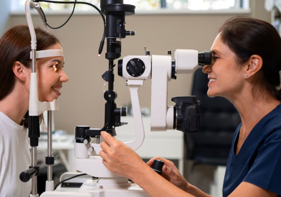
Step 2 – Retinal Examination with BIO
The specialist uses an indirect ophthalmoscope and handheld lens to view the retina in depth, evaluating peripheral and central areas.
Step 3 – Scleral Depression (If Needed)
This technique reveals hidden peripheral tears or retinal elevations.
Step 4 – Documentation and Imaging
Digital recordings, fundus photos, or OCT scans may be taken to confirm findings.
Step 5 – Assessment and Treatment Planning
Clinicians determine if further monitoring, laser treatment, or preventive therapy is required.
Program Duration: 10–20 minutes
Setting: Retinal diagnostic examination room
Recovery & After-Care
After-Care Guidelines
- Expect temporary light sensitivity due to dilation
- Wearing sunglasses is recommended after the exam
- Avoid driving until vision returns to normal clarity
- Report symptoms such as sudden floaters or flashing lights
- Follow the recommended schedule for routine screening
Recovery Timeline
- Immediate: Vision may be blurry for 2–4 hours after dilation
- 1–2 Days: Light sensitivity gradually resolves
- Long-Term: Regular BIO helps detect retinal issues before symptoms develop
Results & Longevity
Expected Results
- Clear visualization of peripheral and posterior retina
- Early detection of retinal tears, lattice degeneration, or detachment
- Accurate assessment of vitreous traction
- Improved risk evaluation for high-myopia patients
- Enhanced safety prior to refractive or cataract surgery
Longevity
- BIO findings remain valuable for long-term monitoring
- Serial exams detect progressive changes in retinal structure
- Retinal risk profiles improve with consistent screening
- Early recognition prevents serious retinal complications
Why Korea Is a Top Destination
- Highly trained retina specialists skilled in indirect ophthalmoscopy
- Integration of BIO with OCT, angiography, and widefield photography
- Advanced diagnostic technology and precise documentation systems
- Exceptional detection accuracy for peripheral retinal diseases
- Pediatric-friendly clinics for newborn and child screening
- Reliable follow-up systems ensuring long-term retinal health
Unique Korean Innovations
- Digital BIO systems with image capture and 3D evaluation
- AI-assisted screening for high-myopia retinal risks
- Advanced scleral depression techniques for ultra-detailed periphery scans
- Structured BIO + OCT + widefield imaging pathways
- Trend tracking for peripheral retinal degeneration
These innovations make Korea’s
Binocular Indirect Ophthalmoscopy Screening Program
one of the most advanced retinal diagnostic screening systems globally.
Cost Range (Indicative Estimate)
| Program Package | Price (KRW) | Approx. USD | Inclusions |
|---|---|---|---|
| Basic BIO Screening | ₩30,000 – ₩70,000 | ~$25 – $55 | Dilated BIO exam |
| BIO + Fundus Photography | ₩70,000 – ₩150,000 | ~$55 – $110 | BIO + digital imaging |
| Comprehensive Retinal Screening Plan | ₩150,000 – ₩350,000+ | ~$110 – $260+ | BIO + OCT + widefield imaging |
Popular Clinics in Korea
- B&VIIT Eye Center (Seoul)
- Dream Eye Center (Seoul)
- BGN Eye Clinic (Seoul & Busan)
- Glory Seoul Eye Clinic
- Kim’s Eye Hospital (Seoul)
- NUNE Eye Hospital (Daegu)
- Seoul National University Hospital Retina Center
- Gangnam Severance Hospital Ophthalmology


