Treatment Overview
Buckle Removal Revision Surgery is a specialized procedure performed to remove a previously placed scleral buckle when it causes complications, discomfort, or structural issues after retinal detachment repair. While scleral buckles are designed to be permanent, certain clinical situations warrant their removal, such as:
- Persistent eye pain
- Extraocular muscle restriction causing double vision
- Buckle infection or exposure
- Migration or extrusion of the buckle
- Chronic conjunctival irritation
- Recurrent redness or inflammation
- Cosmetic concerns
- Interference with future ocular surgeries
In Korea, buckle removal is performed using advanced microsurgical techniques and high-precision imaging systems such as:
- Microscope-assisted buckle dissection
- High-resolution OCT for retinal assessment pre- and post-removal
- Widefield fundus imaging to monitor retina stability
- AI-assisted detection of new retinal breaks or traction risks
- Microincision revision techniques minimizing trauma
Korea’s world-class retinal surgeons ensure safe buckle removal while preserving retinal reattachment and maintaining long-term ocular stability.
Purpose & Benefits
Purpose
- Safely remove a scleral buckle causing symptoms or complications
- Treat buckle-related infections or exposure
- Restore ocular comfort and reduce chronic irritation
- Improve ocular motility and reduce double vision
- Prevent long-term conjunctival scarring or buckle extrusion
- Facilitate future retinal or cataract surgeries
- Assess retinal status and prevent recurrence of detachment
Benefits
- Significant reduction in eye discomfort, redness, and irritation
- Improved eye movement and resolution of diplopia
- Lower risk of buckle migration or infection
- Restored cosmetic appearance
- Minimal risk to retinal stability with modern techniques
- Short surgical time and quick recovery
- Enhanced imaging evaluation before and after removal
Ideal Candidates
Buckle Removal Revision Surgery in Korea is ideal for:
- Patients with buckle exposure or extrusion
- Individuals experiencing chronic eye pain or discomfort
- Those with restricted eye movement due to muscle involvement
- Patients with infection overlying the buckle
- Individuals with persistent redness or conjunctival thinning
- Patients planning cataract or vitrectomy surgeries where buckle interferes
- Anyone needing evaluation for recurrent detachment after buckle placement
Possible Risks & Complications
Buckle removal is safe when performed by retina specialists. However, potential risks include:
Minor/Transient Issues
- Temporary swelling or irritation
- Mild pain after surgery
- Conjunctival redness
- Light sensitivity
Rare/Serious Risks
- Recurrent retinal detachment (rare with proper imaging)
- Conjunctival scarring
- Bleeding around extraocular muscles
- Infection
- Diplopia (typically temporary)
- Retinal traction changes post-removal
Korean clinics minimize risks through:
- Real-time microscope-guided buckle dissection
- Preoperative OCT and widefield imaging to assess retinal security
- Intraoperative inspection for traction changes
- AI-assisted risk analysis for new or potential tears
- Precision suturing with biocompatible materials
Related Diagnostic & Treatment Techniques
- Pars Plana Vitrectomy (if additional revision needed)
- Scleral buckle trimming or partial removal
- Conjunctival reconstruction
- Cryotherapy or laser retinopexy (if new breaks detected)
- Widefield OCT & Fundus Imaging
- B-Scan Ultrasound
- Suture revision for muscle alignment
Treatment Process in Korea
Step 1 – Preoperative Evaluation
- Full eye exam and visual acuity
- Assessment of buckle location and complications
- OCT and widefield imaging to evaluate retina stability
- B-scan ultrasound if visualization is limited
- AI-assisted review for risk of recurrent detachment
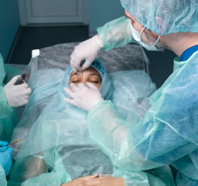
Step 2 – Buckle Removal Surgery
- Local anesthesia with sedation or general anesthesia
- Careful conjunctival opening over the buckle
- Microsurgical dissection to free the buckle
- Removal of sutures and extraction of the buckle material
- Inspection for scleral thinning or muscle involvement
- Conjunctival closure with fine sutures
Step 3 – Optional Revision Procedures
- Cryotherapy or laser if retinal breaks or weak areas detected
- Conjunctival reconstruction for thin or damaged tissue
- Muscle realignment if motility was impaired
Step 4 – Postoperative Follow-Up
- First evaluation within 24–48 hours
- Imaging follow-ups at 1–2 weeks and 1 month
- Long-term monitoring of retinal attachment
Duration: 20–45 minutes
Setting: Specialized retinal surgery suite in a top eye hospital
Recovery & After-Care
After-Care Guidelines
- Use prescribed eye drops (antibiotic + steroid)
- Avoid rubbing or pressing the eye
- Limit heavy lifting for 1–2 weeks
- Keep eye dry for several days
- Avoid strenuous activities
- Attend all scheduled follow-ups
Recovery Timeline
- 24–48 Hours: Reduced discomfort; initial healing
- 1–2 Weeks: Conjunctiva heals; redness improves
- 4–6 Weeks: Normal eye appearance restored
- Long-Term: Stable retinal attachment confirmed
Results & Longevity
Expected Results
- Relief from pain, redness, or irritation
- Improved ocular motility and reduced double vision
- Resolved buckle exposure or infection
- Improved cosmetic appearance
- Stable retina in majority of cases
- Enhanced readiness for future eye procedures
Longevity
- Results are long-lasting as the buckle is permanently removed
- Retina remains stable long-term when pre-op imaging shows no risk
- Follow-up imaging recommended for several months
Why Korea Is a Top Destination
- Exceptional retinal surgical precision
- Highly experienced specialists in buckle revision and complex retinal surgery
- Advanced microscope-integrated imaging systems
- AI-assisted pre- and postoperative retinal evaluation
- Low complication rates with meticulous techniques
- Fast appointment scheduling and efficient surgical pathways
- English-friendly care at major eye centers
Unique Korean Innovations
- AI-enhanced buckle complication analysis
- Ultra-minimally invasive buckle removal techniques
- Customized conjunctival reconstruction for cosmetic restoration
- 3D surgical visualization platforms
- Integrated imaging for long-term retinal surveillance
- Combined buckle removal + vitrectomy protocols for high-risk cases
Cost Range (Indicative Estimate)
| Package | Price (KRW) | Approx. USD | Inclusions |
|---|---|---|---|
| Standard Buckle Removal Surgery | ₩1,200,000 – ₩2,200,000 | ~$900 – $1,700 | Buckle removal + conjunctival repair |
| Advanced Removal with iOCT | ₩2,000,000 – ₩3,800,000 | ~$1,500 – $2,900 | iOCT + widefield imaging + revision care |
| Buckle Removal + Revision Vitrectomy | ₩4,500,000 – ₩8,000,000 | ~$3,450 – $6,150 | PPV + buckle removal + laser/cryotherapy |
| Postoperative Monitoring Package | ₩200,000 – ₩600,000 | ~$150 – $460 | OCT + fundus + AI-assisted analysis |
Popular Clinics in Korea
- Kim’s Eye Hospital (Seoul)
- Gangnam Severance Hospital – Retina Unit
- Seoul National University Hospital Retina Center
- B&VIIT Eye Center (Seoul)
- BGN Eye Clinic (Seoul & Busan)
- NUNE Eye Hospital (Daegu)
- Dream Eye Center (Seoul)
- Glory Seoul Eye Clinic


