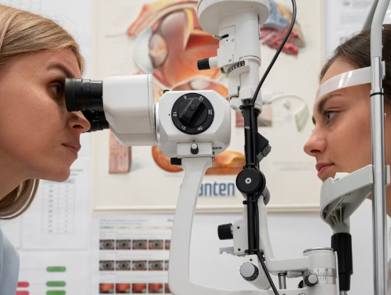
Treatment Overview
BBuckling with Subretinal Fluid Drainage in Korea is a specialized retinal detachment repair technique that combines traditional scleral buckling with controlled removal of subretinal fluid. This approach is used to reattach the retina by:
- Externally supporting the retina with a silicone buckle, and
- Draining accumulated subretinal fluid to allow the retina to flatten and reattach more effectively.
This method is often chosen for cases where retinal detachment includes:
- Large or bullous detachments
- Chronic or long-standing detachment
- Multiple retinal tears
- High fluid accumulation preventing natural reattachment
- Detachments in younger patients, myopic patients, or those with lattice degeneration
Korea is recognized for advanced retinal surgical protocols that integrate:
- High-precision drainage techniques (trans-scleral needle drainage or guarded external drainage)
- Widefield surgical visualization systems (BIOM/Resight)
- High-resolution OCT and ultra-widefield fundus imaging
- Microscope-integrated intraoperative OCT (iOCT)
- AI-assisted mapping of retinal tears and detachment extent
- Minimally invasive scleral buckling materials with improved biocompatibility
These innovations make Korea one of the top destinations for complex or recurrent retinal detachment repair.
Purpose & Benefits
Purpose
- Reattach the retina by externally indenting the sclera
- Drain subretinal fluid to flatten the retina
- Reduce traction on retinal tears
- Provide durable support in cases with significant SRF
- Prevent proliferative vitreoretinopathy (PVR) progression
- Restore normal retinal anatomy and reduce risk of vision loss
Benefits
- Effective for large, chronic, or bullous detachments
- Allows immediate flattening of the retina with drainage
- Minimizes the need for extensive vitrectomy in selected cases
- Long-lasting external support reduces recurrence
- Reduced intraocular manipulation compared to PPV
- Particularly beneficial for young, phakic, and myopic patients
- Can be combined with cryotherapy or laser for optimal sealing
Ideal Candidates
Buckling with SRF Drainage in Korea is ideal for:
- Patients with bullous rhegmatogenous retinal detachment
- Individuals with significant subretinal fluid that prevents natural reattachment
- Young or phakic patients where scleral buckle is preferred
- High myopic patients prone to peripheral retinal tears
- Patients with multiple or difficult-to-access retinal breaks
- Chronic detachments where fluid absorption is limited
- Individuals needing buckle reinforcement without vitrectomy
Possible Risks & Complications
Although safe and widely performed, potential risks include:
Minor/Transient Issues
- Mild postoperative discomfort
- Temporary blurred vision
- Subconjunctival bleeding
- Transient double vision
- Swelling around the eyelid
Rare/Serious Risks
- Infection
- Over- or under-drainage of subretinal fluid
- Choroidal hemorrhage during drainage
- Buckle extrusion (rare with modern materials)
- Recurrent retinal detachment
- Increased myopia due to buckle placement
Korean clinics minimize risks through:
- Controlled needle drainage with real-time visualization
- Intraoperative OCT confirmation
- Precision-calibrated buckle placement
- AI-assisted tear and detachment mapping
- Postoperative monitoring with widefield imaging
Related Diagnostic & Treatment Techniques
- Scleral Buckling Surgery
- Trans-scleral Needle Drainage
- Guarded Subretinal Fluid Drainage
- Cryotherapy for Retinal Tears
- Laser Retinopexy
- Pars Plana Vitrectomy (if needed)
- Gas or Air Tamponade
- Widefield OCT & Fundus Imaging
- B-Scan Ultrasound
Treatment Process in Korea
Step 1 – Preoperative Evaluation
- Complete eye examination and visual acuity
- Widefield retinal imaging (Optos, fundus photography)
- OCT for macular involvement and fluid depth assessment
- Ultrasound B-scan for non-visible areas
- Surgical planning with AI-assisted retinal break detection
Step 2 – Scleral Buckle Placement
- Local or general anesthesia
- Placement of silicone band or sponge on sclera
- Custom buckle height determined for optimal indentation
- Cryotherapy applied to seal retinal tears
Step 3 – Subretinal Fluid Drainage
- Controlled needle or guarded drainage through sclera
- Fluid release to allow the retina to flatten
- Optional air injection to maintain pressure balance
- iOCT used to confirm retinal apposition
Step 4 – Final Adjustments
- Securing the buckle in place
- Ensuring tears are fully sealed
- Confirming retinal reattachment intraoperatively
Step 5 – Postoperative Follow-Up
- First exam within 24 hours
- Subsequent exams at 1 week, 1 month, and ongoing
- OCT and widefield imaging to confirm long-term attachment
Duration: 45–75 minutes
Setting: Advanced retinal surgery room in a specialized eye hospital
Recovery & After-Care
After-Care Guidelines
- Use prescribed eye drops (antibiotics, steroids, anti-inflammatory)
- Avoid heavy lifting and eye pressure
- Keep the operated eye clean and dry
- Avoid rubbing or touching the eye
- Sleep with head elevated for several days
- Attend all postoperative appointments
Recovery Timeline
- 24 Hours: Initial retina stability checked
- 1–2 Weeks: Reduced swelling; early visual recovery
- 1–3 Months: Vision gradually improves
- 3–6 Months: Final retinal healing and stabilization
Results & Longevity
Expected Results
- Successful reattachment of the retina
- Flattening of bullous or chronic detachment areas
- Strong sealing of retinal breaks with buckle support
- Reduced risk of fluid reaccumulation
- Long-lasting structural stability
- Improved visual prognosis, especially when treated early
Longevity
- Scleral buckle provides permanent structural support
- Results last a lifetime unless complicated by new tears or PVR
- Periodic monitoring is recommended for high-risk patients
Why Korea Is a Top Destination
- Leading global success rates for scleral buckling procedures
- Precision drainage techniques minimizing risks
- Advanced iOCT-integrated surgical platforms
- Expert retinal surgeons with high-volume experience
- AI-guided retinal tear detection and surgical planning
- Modernized buckle materials reducing complications
- Comprehensive postoperative monitoring systems
Unique Korean Innovations
- AI-enhanced mapping of subretinal fluid depth
- Guarded drainage cannulas reducing hemorrhage risk
- 3D digital visualization systems for enhanced surgical precision
- Ultra-light silicone buckles reducing induced myopia
- Hybrid techniques combining buckle + vitrectomy when required
- Rapid recovery pathways to restore function sooner
Cost Range (Indicative Estimate)
| Package | Price (KRW) | Approx. USD | Inclusions |
|---|---|---|---|
| Standard Scleral Buckling + SRF Drainage | ₩2,500,000 – ₩4,500,000 | ~$1,900 – $3,450 | Buckle + drainage + cryo/laser |
| Advanced Buckling with iOCT | ₩4,000,000 – ₩6,500,000 | ~$3,050 – $5,000 | iOCT + widefield imaging + premium buckle |
| Combined Buckle + Vitrectomy Package | ₩6,500,000 – ₩10,000,000 | ~$5,000 – $7,650 | PPV + buckle + drainage + gas tamponade |
| Postoperative Imaging Package | ₩300,000 – ₩800,000 | ~$230 – $620 | OCT + fundus + AI analysis |
Popular Clinics in Korea
- Kim’s Eye Hospital (Seoul)
- Gangnam Severance Hospital – Retina Surgery Department
- Seoul National University Hospital Retina Center
- B&VIIT Eye Center (Seoul)
- BGN Eye Clinic (Seoul & Busan)
- NUNE Eye Hospital (Daegu)
- Glory Seoul Eye Clinic
- Dream Eye Center (Seoul)


