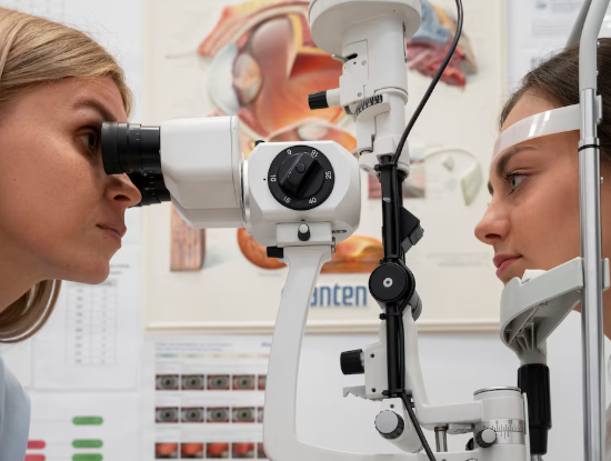
Treatment Overview
EnEndolaser Photocoagulation for Retinal Detachment in Korea is an advanced retinal procedure performed during pars plana vitrectomy (PPV) to seal retinal tears, holes, or areas of weak retina from inside the eye. The procedure delivers focused laser energy directly to the retina through a fiberoptic probe introduced into the vitreous cavity, creating precise chorioretinal scars that promote retinal adhesion.
This method is particularly effective for complex retinal detachments, proliferative vitreoretinopathy (PVR), or detachments associated with vitreous hemorrhage. In Korea, endolaser photocoagulation is performed with cutting-edge technology and highly skilled retinal surgeons:
- High-resolution intraoperative visualization using widefield viewing systems
- Microscope-integrated vitrectomy systems (25–27G PPV)
- Laser probes with adjustable wavelength and power for precise tissue coagulation
- Intraoperative OCT (iOCT) for real-time retinal monitoring
- AI-assisted mapping of retinal breaks and detachment edges
- Integration with adjunctive techniques such as gas or silicone oil tamponade, cryopexy, or scleral buckle if necessary
This approach allows controlled, effective sealing of retinal tissue while minimizing collateral damage and maximizing anatomical success.
Purpose & Benefits
Purpose
- Seal retinal tears, holes, or lattice degeneration during vitrectomy
- Treat rhegmatogenous or complex retinal detachments
- Prevent progression to total detachment or recurrent detachment
- Complement adjunctive procedures like gas/silicone oil tamponade, cryopexy, or scleral buckle
- Reduce vitreoretinal traction and promote retinal stabilization
- Prevent vision loss in complicated retinal detachment cases
Benefits
- High precision and controlled laser delivery inside the eye
- Minimally invasive with microincision vitrectomy
- Effective for complex or posterior retinal breaks
- Can be combined with other repair techniques for maximum success
- Rapid adhesion formation and long-term retinal stability
- Real-time intraoperative monitoring ensures safety
- Reduces risk of postoperative recurrent detachment
Ideal Candidates
Endolaser Photocoagulation in Korea is ideal for:
- Patients with rhegmatogenous retinal detachment
- Individuals with posterior or multiple retinal breaks
- Eyes complicated by vitreous hemorrhage or PVR
- Patients undergoing pars plana vitrectomy for retinal repair
- High-risk patients requiring prophylactic or adjunctive sealing
- Trauma-induced retinal detachment cases
- Individuals with previous retinal surgery needing additional reinforcement
Possible Risks & Complications
Endolaser is generally safe but may have potential risks:
Minor/Transient Issues
- Mild postoperative discomfort or inflammation
- Temporary blurred vision
- Small floaters due to residual vitreous
Rare/Serious Risks
- Incomplete sealing of retinal breaks
- Retinal hemorrhage at laser sites
- Induction of new retinal tears
- Cystoid macular edema or epiretinal membrane formation
- Rare risk of endophthalmitis
- Over- or under-treatment causing localized scotoma
Korean clinics minimize risks through:
- Real-time iOCT monitoring
- Widefield intraoperative visualization
- AI-assisted identification of retinal breaks and detachment areas
- Adjustable laser parameters for energy, duration, and spot size
- Experienced retinal surgeons performing microincision vitrectomy
Related Diagnostic & Treatment Techniques
- Pars Plana Vitrectomy (PPV) – Standard microincision vitrectomy
- Gas or Silicone Oil Tamponade – Maintains retinal reattachment
- Cryopexy or Diathermy – Alternative or adjunctive retinal sealing
- Scleral Buckling – Combined for structural support
- Widefield Fundus Imaging & OCT – Pre- and postoperative evaluation
- AI-Assisted Retinal Mapping – Optimizes laser placement
- Endolaser Fiberoptic Probes – Deliver laser directly inside the vitreous
Treatment Process in Korea
Step 1 – Preoperative Assessment
- Comprehensive eye exam including visual acuity, IOP, and fundus assessment
- OCT and widefield imaging to identify retinal breaks
- AI-assisted analysis to plan laser placement and detachment repair strategy
- Assessment of macula and peripheral retina
Step 2 – Surgical Procedure
- Local or general anesthesia
- 25–27G microincision vitrectomy performed
- Removal of vitreous traction and hemorrhage
- Endolaser applied directly to retinal breaks and surrounding retina
- Gas or silicone oil tamponade injected if required
- Intraoperative OCT ensures complete retinal reattachment and adhesion
Step 3 – Optional Adjunct Procedures
- Cryopexy or diathermy for inaccessible retinal areas
- Scleral buckle placement if structural support is needed
- Fluid–air exchange for optimal retinal flattening
Step 4 – Postoperative Follow-Up
- Immediate postoperative check within 24 hours
- Follow-up imaging at 1 week, 1 month, and 3 months
- OCT and fundus photography to monitor retinal attachment and scar formation
Duration: 60–120 minutes
Setting: Advanced retinal surgery operating room
Recovery & After-Care
After-Care Guidelines
- Use prescribed antibiotic and anti-inflammatory eye drops
- Maintain head positioning if gas tamponade is used
- Avoid strenuous activity, heavy lifting, or bending for several weeks
- Protect the eye from trauma and contact with water or dust
- Attend all scheduled postoperative follow-ups
Recovery Timeline
- Immediate: Blurred vision or gas bubble interference
- 1–2 Weeks: Retina stabilizes; initial vision improvement
- 1–3 Months: Vision improves as gas absorbs and retina adheres
- 3–6 Months: Long-term retinal stability confirmed
Results & Longevity
Expected Results
- Complete sealing of retinal breaks
- Successful retinal reattachment in complex detachments
- Reduced risk of recurrent detachment
- Long-term retinal stability and preservation of vision
- Rapid adhesion formation with minimal postoperative complications
Longevity
- Permanent adhesion at treated sites
- Periodic monitoring recommended for high-risk patients
- Additional intervention rarely needed if procedure is successful
Why Korea Is a Top Destination
- Cutting-edge endolaser systems integrated with vitrectomy microscopes
- AI-assisted preoperative mapping and intraoperative guidance
- Experienced retinal surgeons with extensive vitrectomy expertise
- Combined approach with tamponade, cryopexy, or scleral buckle for complex detachments
- High-volume centers ensure precise and safe treatment
- Efficient outpatient care and follow-up
- English-friendly clinics for international patients
Unique Korean Innovations
- AI-guided intraoperative laser placement for precise sealing
- Widefield visualization for peripheral retinal coverage
- iOCT monitoring for real-time retinal assessment
- Micro-pulse laser delivery to minimize collateral damage
- Hybrid approaches combining laser, cryopexy, and vitrectomy
- Digital surgical tracking for postoperative adhesion and retinal stability
Cost Range (Indicative Estimate)
| Package | Price (KRW) | Approx. USD | Inclusions |
|---|---|---|---|
| Standard Endolaser + PPV | ₩5,000,000 – ₩8,500,000 | ~$3,900 – $6,550 | PPV + endolaser + tamponade + imaging |
| Endolaser + Gas Tamponade | ₩5,500,000 – ₩9,000,000 | ~$4,250 – $6,900 | PPV + endolaser + SF6/C3F8 gas |
| Endolaser + Silicone Oil Tamponade | ₩6,500,000 – ₩10,000,000 | ~$5,000 – $7,650 | PPV + endolaser + silicone oil + imaging |
| Postoperative Monitoring Package | ₩300,000 – ₩800,000 | ~$230 – $620 | OCT + fundus + AI-assisted follow-up |
Popular Clinics in Korea
- Kim’s Eye Hospital (Seoul)
- Gangnam Severance Hospital Retina Unit
- Seoul National University Hospital Retina Center
- B&VIIT Eye Center (Seoul)
- BGN Eye Clinic (Seoul & Busan)
- NUNE Eye Hospital (Daegu)
- Dream Eye Center (Seoul)
- Glory Seoul Eye Clinic


