Treatment Overview
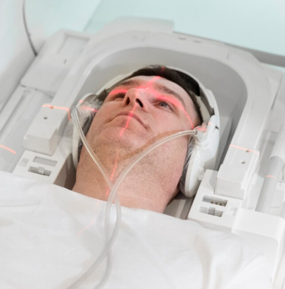
The Fluorescein Angiography (FA) Retinal Scan in Korea is a specialized diagnostic imaging procedure that evaluates blood flow in the retina and choroid using a fluorescent dye. It is primarily used to detect and assess conditions involving retinal blood vessels, macular disorders, and vascular abnormalities.
During the procedure, a safe fluorescent dye (sodium fluorescein) is injected into the bloodstream, and a high-resolution fundus camera captures sequential images as the dye circulates through retinal vessels. This test helps identify leakage, blockages, neovascularization, and ischemic retinal changes with exceptional precision.
Korea is known for its advanced FA retinal scan programs integrating:
- High-speed digital fluorescein angiography systems
- Ultra-widefield (UWF) angiography that captures up to 200° of retina
- AI-assisted vascular analysis for microaneurysm and leakage detection
- Multimodal imaging (FA + OCT + OCTA + FAF) for comprehensive assessment
- Low-allergen, high-purity fluorescein dye for enhanced safety
- Integrated imaging dashboards for detailed disease progression tracking
This diagnostic test is essential for accurate diagnosis, staging, and treatment planning for a wide range of retinal diseases.
Purpose & Benefits
Purpose
- Identify retinal vascular leakage or blockage
- Diagnose diabetic retinopathy, retinal vein occlusions, wet AMD, uveitis
- Assess macular edema and ischemia
- Guide treatment such as laser therapy, anti-VEGF injections, or surgery
- Evaluate abnormalities in retinal or choroidal blood flow
- Confirm progression or stability of existing conditions
Benefits
- Highly detailed imaging of retinal vasculature
- Early detection of vascular abnormalities
- Ultra-widefield imaging detects peripheral lesions otherwise missed
- Assists precise planning for injections or laser procedures
- AI-supported evaluation improves diagnostic accuracy
- Immediate results for same-day treatment decisions
- Essential for detecting hidden leakage or neovascularization
Ideal Candidates
The Fluorescein Angiography Retinal Scan in Korea is suitable for:
- Patients with diabetic retinopathy or suspected microaneurysms
- Individuals with age-related macular degeneration (AMD)
- Patients with retinal vein occlusion (BRVO/CRVO)
- Individuals with unexplained visual distortion or blurred vision
- Patients with uveitis or inflammatory retinal disease
- People with suspected retinal ischemia, leakage, or neovascularization
- Postoperative patients needing vascular assessment
- Patients considered for laser therapy or anti-VEGF injections
Possible Risks & Complications
Fluorescein angiography is generally safe. Potential risks include:
Common/Minor Effects:
- Temporary yellowing of skin and urine (harmless)
- Mild nausea after dye injection
- Temporary blurred vision
- Light sensitivity
Rare/Serious Risks:
- Allergic reaction to fluorescein dye
- Hives or itching
- Very rare anaphylactic reaction
- Vein irritation at the injection site
Korean clinics reduce risks through:
- Allergy screening and preparedness with emergency medications
- High-purity, low-reactive fluorescein dye
- Fast imaging systems that reduce overall exposure
- Skilled medical teams performing all injections and monitoring
Related Diagnostic Techniques
- OCT (Optical Coherence Tomography) – Examines retinal layers
- OCT Angiography (OCTA) – Non-invasive vascular imaging
- Ultra-Widefield Fundus Imaging – Peripheral retina evaluation
- Fundus Autofluorescence (FAF) – RPE abnormalities
- Indocyanine Green (ICG) Angiography – Choroidal vessel assessment
- Slit-Lamp Photography – Anterior segment evaluation
Treatment Process in Korea
Step 1 – Pre-Imaging Assessment
- Vision and IOP check
- Patient history and allergy screening
- Pupil dilation (if needed)
Step 2 – Dye Injection
- A safe fluorescein dye injected into an arm vein
- Patient monitored briefly for any discomfort
Step 3 – Retinal Photography
- High-speed camera captures images as dye travels through retinal vessels
- Sequential phases show arterial, venous, and late leakage patterns
- Optional ultra-widefield scanning to capture peripheral retinal pathology
Step 4 – Specialist Interpretation
- Analysis of leakage, blockage, ischemia, or neovascularization
- Comparison with OCT or previous scans
- Treatment plan recommended based on findings
Step 5 – Follow-Up
- Images stored for long-term comparison
- Repeat FA performed only when needed (e.g., worsening disease)
Duration: 10–20 minutes
Setting: Retinal imaging center or specialized ophthalmology clinic
Recovery & After-Care
After-Care Guidelines
- Drink water to flush dye faster
- Expect yellow-tinged urine for up to 24 hours
- Avoid driving until dilation effects fade
- Notify clinic if severe itching or breathing issues occur
Recovery Timeline
- Immediate: Mild blurred vision and light sensitivity
- 1–2 Hours: Vision normalizes as dilation resolves
- 24 Hours: Fluorescein dye fully eliminated from body
Results & Longevity
Expected Results
- Detailed visualization of retinal blood flow
- Accurate identification of leakage, ischemia, or neovascularization
- Clear guidance for anti-VEGF, steroid injections, or laser treatment
- Reliable tracking of treatment response
Longevity
- Results stored digitally for up to several years
- Serves as baseline for future comparisons
- Repeat scans only needed if disease progression occurs
Why Korea Is a Top Destination
- State-of-the-art angiography cameras and imaging equipment
- Ultra-widefield FA technology for 200° coverage
- Highly trained retina specialists with extensive experience
- AI-integrated diagnostic systems
- Fast, efficient testing with minimal waiting time
- Combined multimodal imaging for the highest diagnostic accuracy
Unique Korean Innovations
- AI-assisted leak detection for early diabetic retinopathy
- Ultra-widefield FA combined with OCTA for hybrid vascular mapping
- High-speed angiography minimizing patient discomfort
- Digital progression dashboards for annual monitoring
- Multimodal imaging suites for one-stop retinal evaluation
These advancements make Korea a global leader in Fluorescein Angiography Retinal Scanning, offering high accuracy and rapid diagnosis essential for advanced retinal care.
Cost Range (Indicative Estimate)
| Package | Price (KRW) | Approx. USD | Inclusions |
|---|---|---|---|
| Standard Fluorescein Angiography | ₩120,000 – ₩250,000 | ~$90 – $190 | FA scan + retinal photography |
| Ultra-Widefield FA Scan | ₩200,000 – ₩350,000 | ~$150 – $270 | Optos widefield FA |
| FA + OCT Package | ₩250,000 – ₩450,000 | ~$190 – $350 | FA + OCT + macular mapping |
| Full Retinal Vascular Panel | ₩300,000 – ₩600,000 | ~$230 – $460 | FA + OCT + OCTA + FAF |
Popular Clinics in Korea
- B&VIIT Eye Center (Seoul)
- Dream Eye Center (Seoul)
- BGN Eye Clinic (Seoul & Busan)
- Kim’s Eye Hospital (Seoul)
- Glory Seoul Eye Clinic
- NUNE Eye Hospital (Daegu)
- Seoul National University Hospital
- Gangnam Severance Hospital Ophthalmology


