Treatment Overview
Fundus Autofluorescence (FAF) Imaging in Korea is a non-invasive diagnostic procedure that captures natural fluorescence from the retinal pigment epithelium (RPE) and other retinal structures. FAF imaging helps detect early retinal changes before they are visible in standard fundus photography or OCT.
FAF is particularly valuable in diagnosing and monitoring age-related macular degeneration (AMD), retinal dystrophies, diabetic retinopathy, central serous chorioretinopathy, and other degenerative retinal conditions.
Korea is internationally recognized for its advanced FAF programs, incorporating:
- High-resolution confocal scanning laser ophthalmoscopy (cSLO)
- Ultra-widefield FAF imaging for peripheral retinal evaluation
- Multimodal imaging integration (FAF + OCT + OCTA)
- AI-assisted autofluorescence pattern analysis for early disease detection
- Sequential imaging dashboards for tracking disease progression
These innovations enable precise mapping of retinal changes, early detection of retinal diseases, and improved treatment planning.
Purpose & Benefits
Purpose
- Detect early retinal pigment epithelium (RPE) abnormalities
- Identify patterns associated with AMD, Stargardt disease, or retinal dystrophies
- Monitor progression of diabetic retinopathy and macular disorders
- Assess photoreceptor and RPE health before clinical symptoms appear
- Guide treatment planning such as anti-VEGF injections or laser therapy
- Track treatment response over time with serial imaging
Benefits
- Non-invasive and painless procedure
- High-resolution imaging of retinal pigment epithelium
- Detects subtle retinal changes invisible on standard fundus photography
- Supports early intervention to prevent vision loss
- AI-assisted analysis improves diagnostic accuracy
- Ultra-widefield imaging identifies peripheral retinal changes
- Rapid acquisition and immediate results for clinical decision-making
Ideal Candidates
FAF Imaging in Korea is recommended for:
- Patients with age-related macular degeneration (AMD)
- Individuals with diabetic retinopathy or suspected retinal ischemia
- Patients with retinal dystrophies (e.g., Stargardt disease)
- Individuals with unexplained visual distortion or central vision changes
- Patients undergoing monitoring of macular edema or degenerative changes
- Postoperative retinal patients needing structural monitoring
- Patients considered for laser therapy or anti-VEGF injections
Possible Risks & Complications
FAF imaging is extremely safe. Potential minor effects include:
Common/Minor Effects:
- Temporary light sensitivity from scanning laser
- Mild eye discomfort during imaging
- Temporary blurred vision if pupils are dilated
Rare/Serious Risks:
- Very rare allergic reaction to dilation drops (if used)
- Minimal risk of dizziness during prolonged imaging
Korean clinics reduce risks through:
- Use of non-dilated imaging when feasible
- Fast, low-intensity scanning laser systems
- Expert technicians ensuring patient comfort
- Pre-screening for light sensitivity or allergies
Related Diagnostic Techniques
- Optical Coherence Tomography (OCT) – Structural assessment of retinal layers
- OCT Angiography (OCTA) – Non-invasive vascular mapping
- Fundus Photography – Color documentation of retina
- Fluorescein Angiography (FA) – Retinal vascular evaluation
- Ultra-Widefield Imaging – Peripheral retina assessment
- Multimodal Retinal Imaging – Integration of FAF, OCT, and OCTA for comprehensive analysis
Diagnostic Process in Korea
Step 1 – Pre-Imaging Assessment
- Review of medical and ocular history
- Visual acuity and intraocular pressure assessment
- Pupil dilation if required
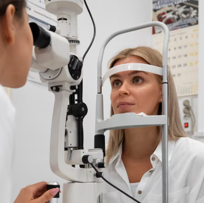
Step 2 – FAF Image Acquisition
- Patient positioned at scanning laser ophthalmoscope
- Sequential images captured showing autofluorescent patterns of RPE
- Optional ultra-widefield imaging for peripheral retina
Step 3 – Specialist Interpretation
- Ophthalmologist analyzes autofluorescence patterns
- Identification of hypo- or hyper-autofluorescent areas indicating retinal stress or disease
- Correlation with OCT/OCTA for comprehensive evaluation
Step 4 – Follow-Up
- Results stored for longitudinal comparison
- Repeat FAF imaging scheduled to monitor progression or treatment response
Duration: 10–20 minutes
Setting: Advanced ophthalmic imaging center
Recovery & After-Care
After-Care Guidelines
- No special recovery required
- Light sensitivity may occur if dilation was used; sunglasses recommended
- Routine daily activities can be resumed immediately
Recovery Timeline
- Immediate: Slight discomfort from bright light exposure
- 1–2 Hours: Vision returns to normal post-dilation
- Long-Term: Periodic FAF imaging provides baseline for disease monitoring
Results & Longevity
Expected Results
- Clear mapping of retinal pigment epithelium health
- Early detection of AMD, dystrophies, or diabetic macular changes
- Precise guidance for treatment planning and intervention
- Documentation for ongoing monitoring and disease management
Longevity
- Images stored digitally for long-term tracking
- Serial imaging enables early detection of progression
- Can be combined with other imaging modalities for comprehensive follow-up
Why Korea Is a Top Destination
- Cutting-edge confocal scanning laser ophthalmoscopes
- Ultra-widefield FAF imaging for full retinal coverage
- AI-assisted analysis for early and accurate detection
- Highly trained retina specialists
- Multimodal imaging suites allowing one-stop diagnosis
- Fast, efficient, and patient-friendly procedures
Unique Korean Innovations
- AI-guided pattern recognition for early AMD detection
- Hybrid FAF + OCT imaging for structural and functional mapping
- Ultra-widefield FAF to capture peripheral retinal changes
- Longitudinal digital dashboards for serial follow-ups
- Integration with multimodal imaging for comprehensive retinal care
These innovations make Korea a leading global center for Fundus Autofluorescence Imaging, offering early detection, precise monitoring, and improved long-term retinal outcomes.
Cost Range (Indicative Estimate)
| Package | Price (KRW) | Approx. USD | Inclusions |
|---|---|---|---|
| Standard FAF Imaging | ₩100,000 – ₩200,000 | ~$75 – $150 | Fundus autofluorescence scan |
| FAF + OCT Package | ₩200,000 – ₩350,000 | ~$150 – $270 | FAF + macular OCT mapping |
| Ultra-Widefield FAF | ₩250,000 – ₩450,000 | ~$190 – $350 | Peripheral retina FAF scan |
| Comprehensive Retinal Panel | ₩300,000 – ₩600,000 | ~$230 – $460 | FAF + OCT + OCTA + multimodal imaging |
Popular Clinics in Korea
- B&VIIT Eye Center (Seoul)
- Dream Eye Center (Seoul)
- BGN Eye Clinic (Seoul & Busan)
- Kim’s Eye Hospital (Seoul)
- Glory Seoul Eye Clinic
- NUNE Eye Hospital (Daegu)
- Seoul National University Hospital Retina Center
- Gangnam Severance Hospital Ophthalmology


