Treatment Overview
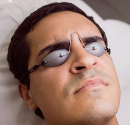
Retinal Break Cryotherapy is a minimally invasive procedure used to seal retinal tears or breaks and prevent progression to retinal detachment. The technique involves applying a precisely controlled cold probe (cryoprobe) to the external sclera overlying the retinal break. This creates a localized adhesion between the retina and the underlying retinal pigment epithelium (RPE), stabilizing the retina.
In Korea, retinal break cryotherapy is performed using advanced imaging, high-precision cryoprobes, and AI-assisted planning:
- Widefield fundus imaging and OCT to locate and map retinal breaks
- Cryotherapy application using modern adjustable probes for precise temperature control
- Combination with laser photocoagulation if needed for complex tears
- Adjunctive use with scleral buckle or vitrectomy in high-risk cases
- AI-assisted monitoring and follow-up for optimal outcomes
Cryotherapy is effective for peripheral retinal tears, lattice degeneration, and retinal holes, and can be performed as a standalone preventive measure or combined with other retinal surgeries.
Purpose & Benefits
Purpose
- Seal retinal breaks and prevent retinal detachment
- Create permanent retinal adhesion through controlled freezing
- Complement other retinal procedures such as scleral buckle or vitrectomy
- Stabilize peripheral retinal tears in high-risk patients
- Provide a minimally invasive alternative to laser photocoagulation when necessary
Benefits
- Minimally invasive outpatient procedure
- High anatomical success rate in preventing detachment from retinal breaks
- Can be combined with vitrectomy or scleral buckle in complex cases
- Rapid treatment with minimal discomfort
- Effective for peripheral retinal tears that are difficult to treat with laser
- AI-assisted planning ensures precise localization and safe application
- Short recovery time with minimal vision interruption
Ideal Candidates
Retinal Break Cryotherapy in Korea is ideal for:
- Patients with peripheral retinal tears or lattice degeneration
- Eyes with rhegmatogenous retinal detachment risk
- High myopia patients prone to retinal breaks
- Individuals with history of trauma or retinal surgery
- Patients where laser photocoagulation is not feasible
- Eyes requiring adjunctive treatment alongside scleral buckle or vitrectomy
Possible Risks & Complications
Minor/Transient Issues
- Mild eye discomfort or irritation at the application site
- Temporary blurred or shimmering vision
- Slight redness or swelling
Rare/Serious Risks
- Recurrent retinal detachment
- Over-freezing causing retinal or choroidal damage
- Vitreous hemorrhage
- Rare scarring or epiretinal membrane formation
- Elevated intraocular pressure (IOP) in susceptible patients
Korean clinics minimize risks through:
- High-resolution imaging for precise retinal break localization
- Modern cryoprobes with adjustable temperature and duration
- Experienced retinal specialists performing the procedure
- AI-assisted planning and follow-up to reduce overtreatment
- Post-procedure monitoring for early detection of complications
Related Diagnostic & Treatment Techniques
- Widefield Fundus Imaging & OCT – Maps retinal breaks and peripheral lesions
- Cryotherapy Application – Creates adhesion between retina and RPE
- Laser Photocoagulation – Complementary or alternative treatment
- Scleral Buckle Surgery – Adjunctive procedure for complex detachments
- Pars Plana Vitrectomy (PPV) – Combined with cryotherapy in high-risk cases
- AI-Assisted Planning & Monitoring – Ensures precise targeting and outcome tracking
Treatment Process in Korea
Step 1 – Preoperative Assessment
- Comprehensive eye examination including visual acuity, IOP, and fundus evaluation
- OCT and widefield fundus imaging to locate retinal breaks
- AI-assisted mapping for optimal cryotherapy application
Step 2 – Surgical Procedure
- Local anesthesia applied
- Cryoprobe placed externally on sclera overlying the retinal break
- Controlled freezing applied until adhesion forms
- Laser photocoagulation may be applied for additional sealing if required
- Post-procedure imaging confirms retinal stability
Step 3 – Postoperative Follow-Up
- Examination within 24–48 hours
- Follow-up imaging at 1 week, 1 month, and 3 months
- Additional cryotherapy or laser applied if new breaks or tears appear
Duration: 20–45 minutes
Setting: Outpatient retinal clinic
Recovery & After-Care
After-Care Guidelines
- Mild discomfort may occur; usually resolves within hours
- Avoid rubbing or pressing the eye for a few days
- Use prescribed anti-inflammatory or antibiotic drops if recommended
- Attend all follow-up imaging sessions
- Report new flashes, floaters, or vision changes immediately
Recovery Timeline
- Immediate: Mild blurred or shimmering vision
- 1–2 Weeks: Retinal adhesion stabilizes; symptoms resolve
- 1–3 Months: Permanent retinal adhesion established
- Long-Term: Monitoring ensures early detection of new breaks
Results & Longevity
Expected Results
- Permanent sealing of retinal breaks
- High success in preventing retinal detachment from treated tears
- Can be combined with other procedures for complex detachments
- Minimal postoperative discomfort and quick return to normal activities
- Optimized outcomes with AI-assisted planning and follow-up
Longevity
- Lifelong retinal monitoring recommended for high-risk patients
- Adhesion from cryotherapy is permanent
- Repeat treatment rarely needed unless new tears develop
Why Korea Is a Top Destination
- Experienced retinal surgeons specializing in cryotherapy
- High-resolution OCT and widefield imaging for precise localization
- AI-assisted planning ensures safe and effective treatment
- Integration with laser, vitrectomy, or scleral buckle if needed
- Efficient outpatient procedure with rapid recovery
- English-friendly clinics for international patients
Unique Korean Innovations
- AI-assisted mapping of retinal breaks for precise cryotherapy application
- Adjustable micro-cryoprobes for controlled freezing
- Widefield imaging for peripheral retinal assessment
- Hybrid treatment protocols combining cryotherapy with laser or vitrectomy
- Digital monitoring dashboards for longitudinal retinal tracking
- Optimized procedural protocols for faster recovery and minimal discomfort
Cost Range (Indicative Estimate)
| Package | Price (KRW) | Approx. USD | Inclusions |
|---|---|---|---|
| Single Retinal Break Cryotherapy Session | ₩500,000 – ₩900,000 | ~$380 – $700 | Cryotherapy + imaging + AI-assisted planning |
| Cryotherapy + Laser Combination | ₩800,000 – ₩1,500,000 | ~$620 – $1,150 | Dual treatment for complex retinal breaks |
| Follow-Up Monitoring Package | ₩300,000 – ₩800,000 | ~$230 – $620 | 1–3 follow-up OCT/fundus sessions |
Popular Clinics in Korea
- Kim’s Eye Hospital (Seoul)
- Gangnam Severance Hospital Retina Unit
- Seoul National University Hospital Retina Center
- B&VIIT Eye Center (Seoul)
- BGN Eye Clinic (Seoul & Busan)
- Dream Eye Center (Seoul)
- NUNE Eye Hospital (Daegu)
- Glory Seoul Eye Clinic


