Treatment Overview
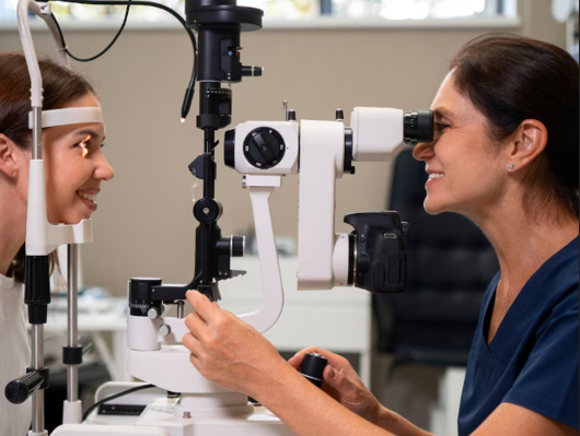
Retinal Neovascularization Monitoring is a diagnostic and follow-up program designed to detect and track the growth of abnormal blood vessels in the retina. Neovascularization is a hallmark of proliferative diabetic retinopathy (PDR), retinal vein occlusion, and other ischemic retinal diseases, and can lead to vitreous hemorrhage, tractional retinal detachment, and severe vision loss.
Korea is internationally recognized for advanced monitoring programs that integrate:
- High-resolution Optical Coherence Tomography Angiography (OCTA) for non-invasive vascular imaging
- Fluorescein Angiography (FA) and Indocyanine Green Angiography (ICG) for perfusion and leakage assessment
- Fundus photography and fundus autofluorescence (FAF) for structural and pigment evaluation
- AI-assisted detection and quantification of neovascular growth
- Integration with intravitreal therapy or laser treatment planning
- Rapid outpatient monitoring with serial imaging for disease progression tracking
This program enables early detection, timely intervention, and precise management of retinal neovascularization, minimizing the risk of vision-threatening complications.
Purpose & Benefits
Purpose
- Detect early formation of abnormal retinal vessels
- Monitor progression of neovascularization in diabetic or ischemic retinal disease
- Evaluate treatment response after anti-VEGF injections, laser therapy, or vitrectomy
- Guide clinical decisions for timely intervention
- Prevent complications like vitreous hemorrhage and tractional retinal detachment
- Provide baseline and longitudinal data for chronic retinal conditions
Benefits
- Non-invasive and minimally discomforting procedures
- High-resolution visualization of retinal and choroidal vasculature
- AI-assisted early detection of subtle microvascular changes
- Real-time monitoring enables timely therapeutic interventions
- Reduced risk of vision loss due to proactive management
- Can be combined with OCT, FA, ICG, and fundus imaging for comprehensive assessment
- Supports personalized treatment planning and follow-up
Ideal Candidates
Retinal Neovascularization Monitoring in Korea is ideal for:
- Patients with proliferative diabetic retinopathy (PDR)
- Individuals with ischemic retinal vein occlusion
- Patients with age-related macular degeneration (AMD) with neovascularization risk
- Individuals with history of vitreous hemorrhage or retinal detachment
- High-risk diabetic or hypertensive patients requiring regular retinal surveillance
- Patients undergoing anti-VEGF therapy, laser therapy, or retinal surgery
Possible Risks & Complications
Monitoring procedures are generally safe and non-invasive. Potential considerations include:
Minor/Transient Issues
- Mild eye strain during imaging
- Difficulty maintaining fixation in uncooperative patients
- Motion artifacts affecting image clarity
Rare/Minimal Risks
- Allergic reaction to fluorescein or indocyanine green dye (for FA/ICG)
- Temporary transient visual disturbances post-dye injection
Korean clinics minimize risks through:
- Experienced technicians performing imaging
- Motion artifact correction software
- Pre-screening for dye allergies
- AI-assisted analysis to reduce repeated scans
Related Diagnostic & Treatment Techniques
- OCTA (Optical Coherence Tomography Angiography) – Non-invasive vascular mapping
- Fluorescein Angiography (FA) – Detects leakage and perfusion defects
- Indocyanine Green Angiography (ICG) – Evaluates choroidal neovascularization
- Fundus Photography & FAF – Tracks structural retinal changes
- Intravitreal Anti-VEGF Therapy – Guided by neovascularization monitoring
- Laser Therapy (PRP, Micro-Pulse, Barricade) – Targeted based on neovascular maps
- Pars Plana Vitrectomy – For hemorrhage or traction associated with neovascularization
Treatment Process in Korea
Step 1 – Baseline Assessment
- Comprehensive eye exam including visual acuity, IOP, and fundus evaluation
- OCT and OCTA imaging to identify retinal and choroidal vessels
- Fluorescein or ICG angiography if indicated
Step 2 – AI-Assisted Analysis
- Automated detection and quantification of neovascular growth
- Comparison with prior images for longitudinal assessment
- Mapping of ischemic zones and leakage areas
Step 3 – Clinical Correlation
- Review by retinal specialists to determine progression
- Planning interventions such as anti-VEGF therapy or laser treatment
- Integration with multimodal imaging for personalized care
Step 4 – Follow-Up Monitoring
- Regular OCTA, OCT, and FA imaging intervals (typically 3–12 months, depending on risk)
- Adjustment of treatment protocols based on neovascular activity
- Early intervention to prevent hemorrhage, detachment, or vision loss
Duration: 15–45 minutes per session
Setting: Advanced retinal imaging center or ophthalmology clinic
Recovery & After-Care
After-Care Guidelines
- No downtime; patients can resume normal activities immediately
- Monitor for any allergic reactions if dye angiography is performed
- Attend scheduled follow-up imaging sessions
- Maintain systemic disease control (diabetes, hypertension) to reduce neovascular risk
Recovery Timeline
- Immediate: Imaging results are available immediately or within hours
- Short-Term: Clinical evaluation guides interventions if needed
- Long-Term: Serial monitoring ensures early detection of recurrent or progressive neovascularization
Results & Longevity
Expected Results
- Early detection and accurate monitoring of retinal neovascularization
- Quantitative mapping of vessel density, leakage, and ischemic areas
- Optimized timing for intravitreal therapy or laser treatment
- Reduced risk of vitreous hemorrhage, retinal detachment, and vision loss
- Personalized care with long-term disease management
Longevity
- Lifelong monitoring recommended for high-risk patients
- Non-invasive imaging allows repeated evaluation without cumulative risk
- AI-assisted longitudinal tracking enhances early intervention and outcome prediction
Why Korea Is a Top Destination
- Advanced OCTA, OCT, FA, and ICG imaging systems
- AI-assisted retinal vascular analysis for early detection
- Experienced retinal specialists for interpretation and treatment planning
- Integration with multimodal imaging and intravitreal therapy
- Efficient outpatient monitoring with rapid, accurate results
- Personalized follow-up schedules for chronic retinal disease
Unique Korean Innovations
- AI-assisted neovascular detection and progression analysis
- Widefield OCTA and FA for peripheral retina assessment
- Quantitative vessel density mapping for treatment optimization
- Integration with intravitreal anti-VEGF therapy planning
- Digital dashboards for longitudinal monitoring and patient management
Cost Range (Indicative Estimate)
| Package | Price (KRW) | Approx. USD | Inclusions |
|---|---|---|---|
| Single OCTA Scan | ₩200,000 – ₩400,000 | ~$150 – $310 | Non-invasive vascular imaging + AI-assisted report |
| OCT + OCTA Comprehensive Assessment | ₩500,000 – ₩900,000 | ~$380 – $700 | Structural OCT + angiography + interpretation |
| FA or ICG Dye Angiography | ₩400,000 – ₩800,000 | ~$310 – $620 | Dye-based imaging + analysis |
| Serial Monitoring Package | ₩900,000 – ₩1,500,000 | ~$700 – $1,150 | 3–5 imaging sessions over 3–6 months + AI-assisted analysis |
Popular Clinics in Korea
- B&VIIT Eye Center (Seoul)
- Dream Eye Center (Seoul)
- BGN Eye Clinic (Seoul & Busan)
- Kim’s Eye Hospital (Seoul)
- Seoul National University Hospital Retina Center
- NUNE Eye Hospital (Daegu)
- Glory Seoul Eye Clinic
- Gangnam Severance Hospital Ophthalmology


